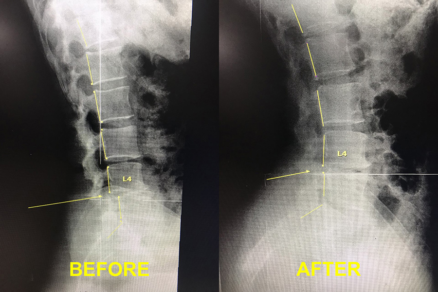
A normal lumbar curve should approximate an elliptical curve and the back of each lumbar should be lined up. When injuries occur the lumbar curve is often reduced and slipping of the vertebrae can occur. This can cause low back pain, muscle spam, sciatica, leg pain, hip pain, weakness and numbness into the lower extremity. Each patient will present with unique injury patterns that can only be detected with weight bearing x ray imaging. After proper x ray analysis is performed a correction plan can be implemented. This example demonstrates a loss of the lumbar lordosis with sliding of the L4 vertebrae and the correction after a series of adjustments and lumbar traction sessions were administered.
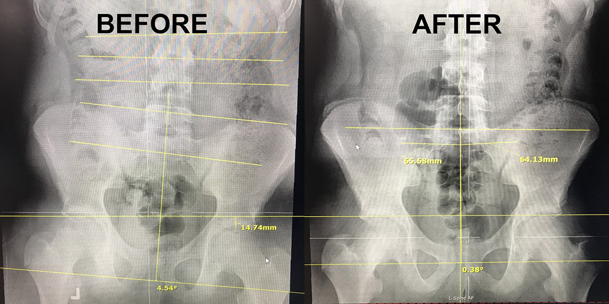
Due to anomalies in the growth cycle one side of the body may grow larger than the other causing a structural shift to the pelvis. Many people with chronic low back and hip pain have a structural issue that creates a functional movement disorder that leads to chronic subluxation patterns. These patterns cannot be corrected using chiropractic adjustments alone. If a structural issue is suspected proper x ray placement and analysis is utilized to correct for the structural inequality. This allows the patient to maintain proper alignment and biomechanics allowing the spine to function properly.
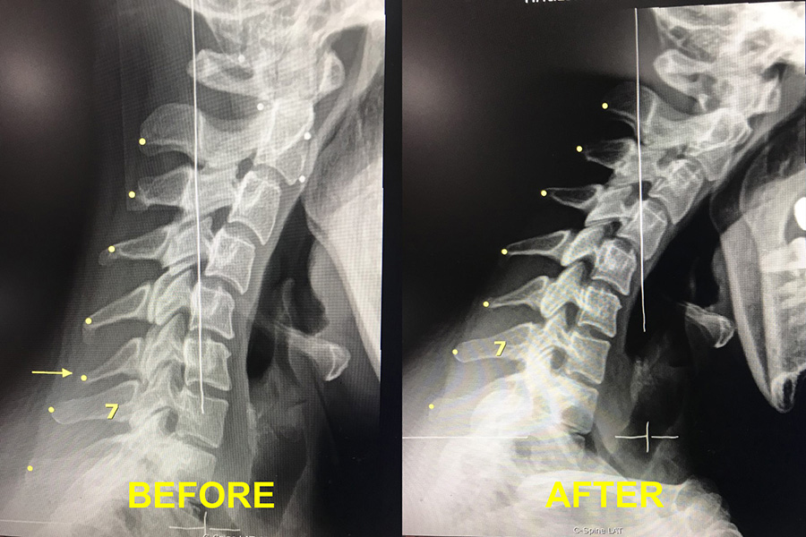
The cervical flexion/extension x ray is one of the most valuable tools in identifying subluxated vertebrae in the cervical spine or neck. In normal flexion the vertebrae should tilt forward and the back portion called the spinous process should fan open. In this example the C6 vertebrae is stuck in extension even when the patient is in full flexion. Upon identifying the subluxated vertebrae a correction can be made and then verified that full restoration of function has been made after a series of adjustments. Chronic neck pain, shoulder pain, muscle spam, numbness and tingling down the arms and headache can be contributed to cervical subluxation.
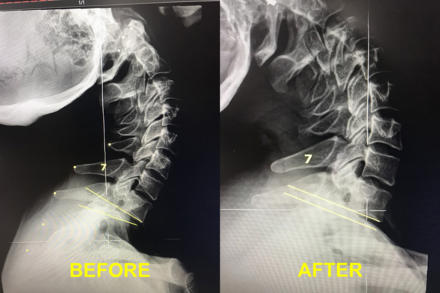
The cervical flexion/extension x ray is one of the most valuable tools in identifying subluxated vertebrae in the cervical spine or neck. In normal extension the vertebrae should tilt backward and the back portion called the spinous process should close down and approximate. In this example the T1 vertebrae is stuck in flexion even when the patient is in full extension. Upon identifying the subluxated vertebrae a correction can be made and then verified that full restoration of function has been made after a series of adjustments. Chronic neck pain, shoulder pain, muscle spam, numbness and tingling down the arms and headache can be contributed to cervical subluxation.
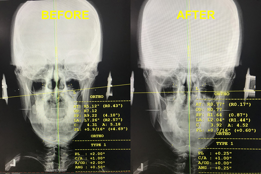
The uppercervical spine consists of the base of the skull (Occiput) the Atlas and the Axis vertebrae. In the absence of malformation the properly aligned Atlas will sit level relative to the horizon; the head and cervical spine are vertical and perpendicular to the atlas vertebrae. The spinous processes of the cervical vertebrae should be lined up in the center of the vertebrae with no twist or rotation. When the neck and head are in perfect alignment the brainstem and spinal cord can sit in the center of the spinal canal free of interference from torque produced by pulling on ligamentous attachments called the dentate ligaments. The Atlas is the most freely movable vertebrae in the spine with half of normal head rotation coming from the C1/2 articulation. Chronic malposition and subluxation of the Atlas vertebrae can cause insufficiency in blood flow of the vertebral artery and the jugular vein, congestion of cerebral spinal fluid may also occur along with torsion to the cerebellum and brainstem. These effects may result in different types of headaches, neurological symptoms, balance disorders, dizziness and unsteadiness, face pain, jaw pain, neck and shoulder pain, lower spine and pelvis misalignment and many other symptoms. Proper uppercervical analysis and correction is crucial for full healing to occur and for the central nervous system to communicate free of interference.
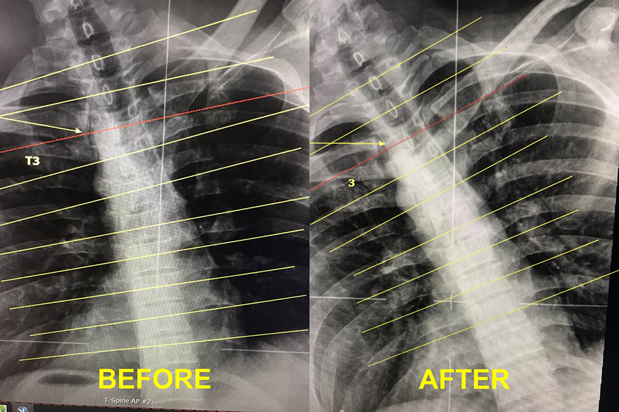
The motion analysis x ray is one of the most valuable tools in subluxation diagnostic analysis to locate difficult to find vertebral subluxations. The spine has a normal movement pattern unique to each region. In this left lateral bending x ray the vertebrae should close on the left and open on the right. The T3 vertebrae demonstrated by the red line is stuck bending to the right even though the patient is bending to the left. Upon identifying the subluxated vertebrae a correction can be made and then verified that full restoration of function has been made after a series of adjustments. Certain patients with chronic subluxation patterns may need a very detailed analysis to find a subluxation often missed by palpation or a standard neutral x ray. The motion x ray analysis allows the detail oriented chiropractor to identify hidden subluxations that may otherwise go undetected.